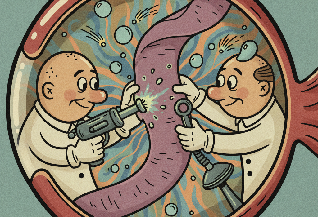
Imagine the inside of your eye is like a room lined with delicate, light-sensitive wallpaper. This “wallpaper” is your retina, and its job is to capture light and send images to your brain. A retinal detachment happens when this tissue peels away from the back wall of the eye.
When the retina detaches, it loses its blood supply and stops working, which can lead to permanent vision loss if not treated quickly. For decades, researchers have worked to find the safest and most effective ways to press this vital tissue back into place.
Today, treating a retinal detachment is not a one-size-fits-all process. The right treatment depends entirely on how and why the retina detached. In this article, we will explore what peer-reviewed research says about the different types of retinal detachments, how modern treatments work, and who benefits most from specific procedures.
Understanding the Three Types of Detachment
To understand the treatments, we first need to understand the three main ways a retina can detach. Science categorizes them based on the underlying cause:
1. Rhegmatogenous (reg-mah-TOJ-uh-nus): A tear or hole allows fluid to leak behind the retina, pushing it off the wall.
2. Tractional: Scar tissue forms on the retina’s surface and physically pulls it away.
3. Exudative (ex-YOO-day-tiv): Fluid builds up beneath the retina due to inflammation or vascular disease, without any tears being present.
What the Research Shows About Treatment Options
Doctors have several tools to reattach the retina. The choice depends heavily on the type of detachment and the patient’s specific anatomy.
Pneumatic Retinopexy: The Gas Bubble Technique
For many rhegmatogenous detachments (those caused by a tear), doctors use a procedure called pneumatic retinopexy (noo-MAT-ik RET-in-oh-pek-see). A doctor injects a small gas bubble into the eye. The patient then holds their head in a specific position so the bubble floats up and presses the detached retina back into place. Once the retina is flat, a laser or freezing tool is used to “weld” the tear shut.
Historically, this was only used for tears at the top of the eye, relying on gravity to make the bubble float upward. However, a 2011 study in Retina found that this less invasive, outpatient technique can also work for tears at the bottom of the eye. By having patients lie on their side with their head tilted downward at a 30 to 60-degree angle, the gas bubble successfully pressed against the bottom tears, achieving a 76.9% initial success rate without the need for major surgery.
Early research, including a 1989 study from South Africa, confirmed that using these inert gases allows many patients to be treated in an outpatient setting, avoiding lengthy hospital stays.
Vitrectomy and Physical Tamponades
When a detachment is complex, doctors may perform a vitrectomy (vih-TREK-tuh-mee). This involves removing the jelly-like substance inside the eye and replacing it with a temporary filler, or tamponade, to hold the retina flat.
Fillers usually include expanding gases or silicone oil. A 1986 study in The British Journal of Ophthalmology looked at patients with severe scar tissue pulling on the retina. The researchers found that using extended fillers like sulfur hexafluoride gas or silicone oil resulted in a 68% success rate for reattachment. Silicone oil is often preferred for patients who cannot maintain the strict head positioning required for gas bubbles.
Scientists are also testing new materials. A 2021 animal study in Current Eye Research tested a new hyaluronic acid hydrogel as a possible alternative to gas and oil. While it showed excellent ability to hold the retina in place, some subjects developed inflammation, meaning more research is needed before it becomes standard for humans.
Treating Exudative Detachments (Fluid Buildup)
Exudative detachments are tricky because there is no tear to seal. Instead, doctors must treat the underlying disease causing the fluid leak.
For example, highly nearsighted people sometimes develop a condition called “dome-shaped macula,” which can cause fluid to pool under the center of the retina. A 2018 pilot study in Retina tested a gentle laser therapy called subthreshold laser treatment on these patients. After 12 months, the patients showed significant improvements in visual acuity and a reduction in fluid thickness, even if the fluid did not disappear completely.
In rare cases, severe inflammation can cause fluid buildup. A 2021 case report in the European Journal of Ophthalmology highlighted how an undiagnosed infection (ocular syphilis) caused an exudative detachment when mistakenly treated with steroids alone. Once the underlying infection was treated with antibiotics, the detachment resolved, showing how critical accurate diagnosis is for this specific type of detachment.
Related: How to Prevent Diabetic Eye Disease: What the Science Says
Who Benefits Most From Specific Approaches
Research clearly identifies certain populations that require highly tailored approaches to retinal detachment.
Premature Infants
Babies born very early can develop Retinopathy of Prematurity (ROP), where abnormal blood vessels grow and create scar tissue that pulls the retina off the wall (tractional detachment).
Doctors debate whether to use laser therapy to stop the vessel growth or inject an anti-VEGF medication (which blocks the signal causing the abnormal vessels). Two major studies provide clarity on this:
- A 2021 study in Ophthalmology analyzed 1,167 eyes of premature infants.
- A 2019 study in the Journal of AAPOS looked at 222 eyes.
Both studies found a clear dividing line based on the infant’s age.
| Infant Age at Treatment | Anti-VEGF Injection | Laser Therapy | Which is Better? |
|---|---|---|---|
| Before 36 weeks | 0% detachment rate | 7.9% – 16% detachment rate | Anti-VEGF is significantly better at preventing short-term detachment. |
| After 36 weeks | 1.4% detachment rate | 0.8% – 3.1% detachment rate | Both work equally well; no statistical difference. |
Because anti-VEGF medications work faster than lasers, they are much better at halting aggressive disease in the youngest, most vulnerable infants.
Related: Eye Injections for Macular Degeneration: What Science Says
Protecting the “Good” Eye
If you have a retinal tear in one eye, what are the chances it will happen to the other? Research shows the risk is notable, prompting doctors to investigate preventative laser treatments.
A 1990 study in France analyzed 302 “second” eyes of patients who had already suffered a detachment in their first eye. The researchers applied a preventative argon laser treatment around the edges of the healthy retina. They found that this systematic prophylactic treatment decreased the rate of a second detachment from 11% down to just 2.4%. A follow-up 1991 study confirmed these long-term benefits, showing that securing weak spots with a laser is highly effective for high-risk patients.
Where The Science Is Still Evolving
While standard detachments have high success rates, some anatomical anomalies remain difficult to treat.
For instance, an optic nerve head coloboma is a rare birth defect where tissue is missing from the optic nerve, sometimes leading to a detached retina. A 2012 review noted that these are notoriously difficult to repair because standard lasers cannot be safely applied to the optic nerve. Researchers are experimenting with novel techniques, such as using a patient’s own blood platelets to create a natural clot to seal the hole, but this science is still in its early stages.
Additionally, treating eye tumors can occasionally cause the retina to detach. A 2008 study in Retina found that about 1% of patients treated with heat therapy (thermotherapy) for choroidal melanoma developed a retinal break. Fortunately, these could usually be successfully repaired with standard vitrectomy surgery.
Common Questions About Retinal Detachment
Do I have to lay face down after surgery?
If your doctor uses a gas bubble to push the retina into place, you will likely need to maintain a specific head position. While this historically meant lying face down, newer research shows that lying on your side with your head tilted can work for tears in the lower part of the eye.
Can a detachment heal on its own?
No. A rhegmatogenous or tractional retinal detachment is a mechanical problem that requires a mechanical fix (like a gas bubble, laser, or surgery). Without treatment, it will lead to permanent blindness.
Does laser treatment hurt?
Most patients report mild discomfort or a sensation of light pressure during preventative laser treatments, but it is generally well-tolerated and performed in an outpatient setting.
The Bottom Line
Retinal detachment is a serious medical emergency, but modern science offers highly effective treatments. The evidence shows:
- Pneumatic retinopexy (gas bubbles) is a highly successful, less invasive option for many tears, even those at the bottom of the eye if proper head positioning is used.
- Vitrectomy with gas or silicone oil remains the gold standard for complex detachments involving scar tissue.
- Anti-VEGF injections provide superior short-term protection against detachment for very premature infants (under 36 weeks) compared to traditional lasers.
- Preventative laser therapy on a healthy eye can significantly reduce the risk of a future detachment if the other eye has already suffered one.
If you experience sudden flashes of light, a shower of new floaters, or a dark curtain falling over your vision, seek immediate emergency eye care. Time is the most critical factor in saving your sight.
Quick Reference: Key Studies
| Study Focus | Key Finding | Source |
|---|---|---|
| Premature Infants (ROP) | Anti-VEGF injections prevent short-term detachment better than lasers in infants under 36 weeks. | PMID 33387554 & PMID 31513902 |
| Pneumatic Retinopexy | Gas bubble therapy is effective for lower-eye retinal tears when patients use a tilted side-lying posture. | PMID 21052037 |
| Preventative Laser | Prophylactic laser treatment on a healthy eye reduces the risk of bilateral detachment from 11% to 2.4%. | PMID 2192808 |
| Severe Scarring (PVR) | Extended use of gas or silicone oil fillers during surgery yields a 68% reattachment success rate. | PMID 3801368 |
| Fluid Buildup (Exudative) | Subthreshold laser treatment safely reduces fluid and improves vision in highly nearsighted eyes. | PMID 28145971 |
Last updated: March 2026
This article synthesizes findings from peer-reviewed research. It is for educational purposes only and does not constitute medical advice. Consult a healthcare provider before starting any new regimen.
Leave a Reply