Understanding the Pressure Inside Your Eye
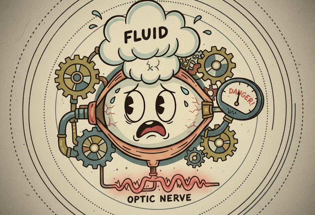
Imagine your eye is like a sink with the faucet always running. To keep the water level balanced, the sink has a drain. In your eye, a clear fluid called aqueous humor constantly flows in to provide nutrients. It then drains out through a spongy tissue near the edge of your cornea.

If that drain gets clogged, fluid builds up. This increases your Intraocular pressure (in-trah-AHK-yoo-ler PRESH-ur), which is the fluid pressure inside your eye. Over time, high pressure can crush the delicate optic nerve at the back of your eye. This nerve is the cable that sends images to your brain. When it gets damaged, blind spots develop. This condition is known as glaucoma.
For decades, doctors believed that high eye pressure was the sole cause of glaucoma. However, modern science paints a much more complex picture. Many people with high eye pressure never develop nerve damage. Even more surprisingly, some people develop severe glaucoma while their eye pressure remains completely normal.
Researchers now know that glaucoma is not just a plumbing problem. It involves blood flow, brain fluid pressure, cellular stress, and even your daily physical activities.
Related: How to Prevent Diabetic Eye Disease: What the Science Says
How This Might Work: The Tug-of-War Behind Your Eye
To understand why normal eye pressure can still cause blindness, we have to look at the exact spot where the optic nerve leaves the eye. This exit point is called the Lamina cribrosa (LAM-ih-nuh crib-ROH-suh). It is a mesh-like wall that allows nerve fibers to pass through.
This wall sits right between two different pressurized zones. Inside your eye, the intraocular pressure pushes outward against the wall. Behind your eye, the fluid that surrounds your brain and spinal cord pushes inward. This brain fluid has its own pressure, known as Cerebrospinal fluid pressure (seh-ree-bro-SPY-nul FLOO-id PRESH-ur).
Scientists call the balance between these two forces the trans-lamina cribrosa pressure difference. It is a constant tug-of-war.
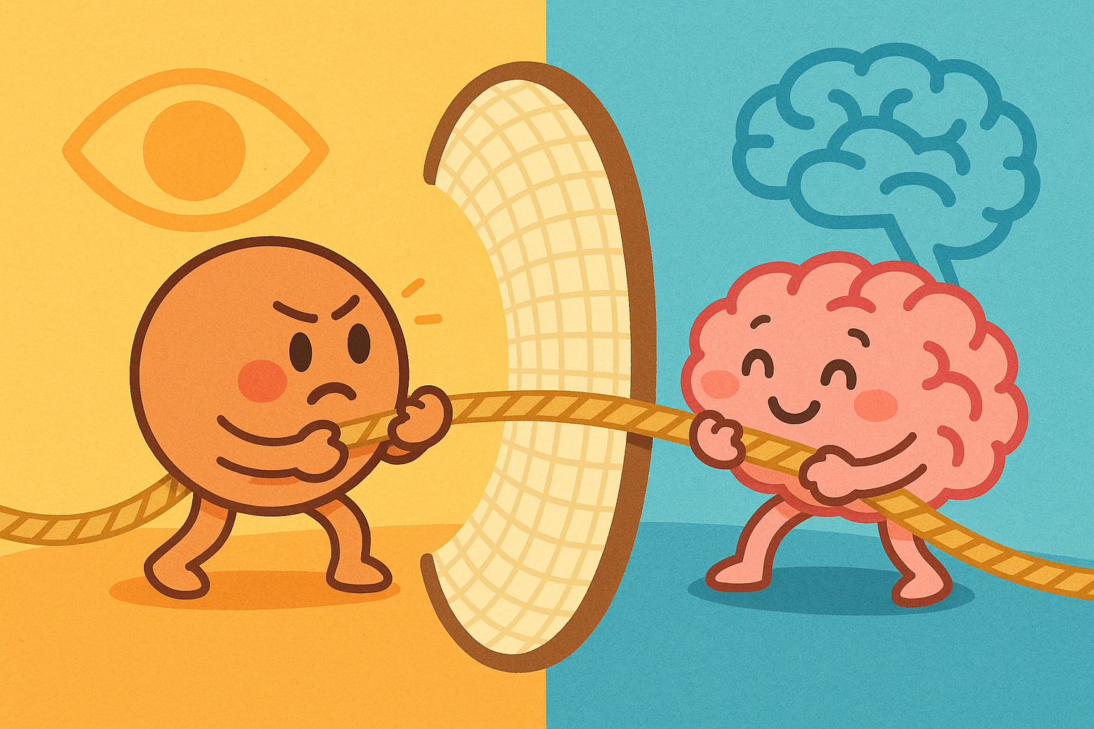
A 2014 review in The British journal of ophthalmology explains that if your brain fluid pressure is unusually low, the inward push is very weak. As a result, even a “normal” amount of eye pressure can easily overpower the brain fluid pressure. The wall bows outward, stretching and damaging the optic nerve fibers. This helps explain why people develop normal-tension glaucoma.
What the Research Shows
Recent studies have uncovered several hidden factors that drive glaucoma progression. The research highlights that pressure spikes, blood flow, and cellular rust all play major roles.
Hidden Pressure Spikes in Daily Life
When you visit the eye doctor, they measure your pressure in a calm, seated position. But your eye pressure does not stay the same all day. It fluctuates constantly.
A study in Optometry and vision science found that simple daily activities can cause massive, temporary spikes in eye pressure.
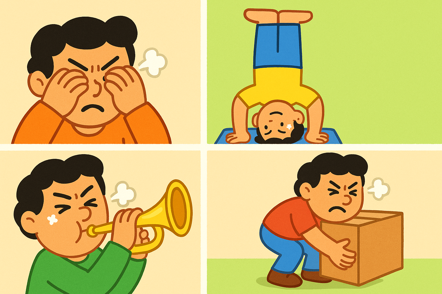
Some common triggers include:
- Eye rubbing: Vigorously rubbing your eyes can temporarily raise pressure by hundreds of millimeters of mercury.
- Inverted postures: Yoga headstands or using gravity boots cause blood and fluid to rush to the head, drastically raising eye pressure.
- Wind instruments: Playing high-resistance instruments like the trumpet or oboe increases pressure inside the chest, which limits blood return from the head and spikes eye pressure.
- Heavy lifting: Straining while holding your breath forces pressure upward into the eyes.
For a healthy eye, these brief spikes are harmless. But for an eye that is already vulnerable, repeated pressure spikes can stretch the tissues and accelerate nerve damage over time.
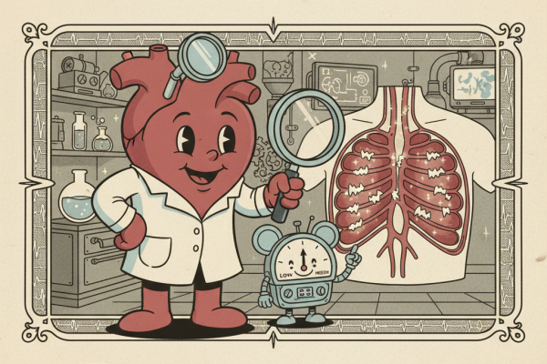
Blood Flow and Vascular Health
Your optic nerve needs a steady supply of oxygen-rich blood to survive. If blood flow is unstable, the nerve fibers can starve.
Researchers in the American journal of ophthalmology studied patients whose glaucoma continued to worsen even after their eye pressure was medically lowered. They found that these patients often had poor blood flow behind the eye.
This is known as vascular dysregulation. It means the blood vessels do not widen and narrow properly in response to the body’s needs. People with this issue often have low systemic blood pressure or suffer from cold hands and feet. When blood pressure drops too low at night, the optic nerve does not get enough oxygen, leading to slow, progressive damage.
Cellular Rust and Oxidative Stress
When cells in your eye are stressed by high pressure or poor blood flow, they produce Reactive oxygen species (ree-AK-tiv OX-ee-jen SPEE-sheez). These are unstable molecules that bounce around and damage healthy cells. You can think of this process as cellular rust, formally known as oxidative stress.
A review in Progress in brain research shows that oxidative stress heavily damages the Trabecular meshwork (tra-BEK-yoo-ler MESH-werk). This is the primary drain of the eye. As the cells rust and die, the drain becomes stiff and clogged with debris. This creates a vicious cycle. The clogged drain raises eye pressure, which creates more oxidative stress, which further damages the drain.
Secondary Causes of Glaucoma
Sometimes, glaucoma is a secondary side effect of another distinct eye problem.
- Eye Cancer: Tumors inside the eye, like melanoma, can physically push the lens forward and block the drain. In other cases, the tumor sheds cells and debris that physically clog the meshwork. A review in Survey of ophthalmology highlights that a sudden, severe case of glaucoma in just one eye can sometimes be the first sign of an underlying tumor.
- Surgical Gels: During cataract surgery, doctors use thick, clear gels called viscoelastics to protect the inside of the eye. A study in Seminars in ophthalmology notes that if these large gel molecules are not completely washed out after surgery, they can temporarily plug the eye’s drain and cause severe pressure spikes.
- Malignant Glaucoma: This is a rare, severe complication that can happen after eye surgery. Fluid gets misdirected backward into the jelly-like center of the eye. This pushes everything forward, completely pancaking the front of the eye. A historical review in Survey of ophthalmology explains that traditional glaucoma drops actually make this specific condition worse, requiring special medications or surgery to fix.
Who Benefits Or Needs Caution
Research clearly identifies populations that face different risks when it comes to glaucoma.
- Asian Populations: A 2023 study in International ophthalmology found that Asian populations represent a massive portion of global glaucoma cases. They are uniquely prone to angle-closure glaucoma and normal-tension glaucoma.
- Women: The same global study found that women comprise the majority of all glaucoma cases worldwide.
- Highly Nearsighted Individuals: People with severe myopia have elongated eyes. This physical stretching weakens the tissues around the optic nerve, making them more vulnerable to pressure damage.
- Young Adults: While rare, juvenile-onset open-angle glaucoma can strike people under 40. A 2022 review in Survey of ophthalmology notes this is heavily tied to genetics and often presents with very high eye pressures.
Comparing Glaucoma Types
| Feature | High-Tension Glaucoma | Normal-Tension Glaucoma |
|---|---|---|
| Eye Pressure | Consistently high (above 21 mm Hg) | Statistically normal (below 21 mm Hg) |
| Primary Cause | Clogged drainage system | Poor blood flow or low brain fluid pressure |
| Associated Risks | Age, family history, thin corneas | Migraines, cold hands, low blood pressure |
| Treatment Focus | Lowering eye pressure | Lowering eye pressure and supporting blood flow |
Where The Science Is Still Uncertain
Because eye pressure fluctuates, doctors have long wondered if monitoring a patient’s pressure for a full 24 hours would help predict if their disease will get worse.
However, a study in Graefe’s archive for clinical and experimental ophthalmology found that 24-hour pressure monitoring in a hospital setting was surprisingly ineffective. The pressure spikes recorded during the hospital stay did not accurately predict which patients would lose more vision. The researchers concluded that single-day monitoring in an artificial environment is a poor diagnostic tool, highlighting the need for better wearable sensors in the future.
Practical Guidance and Treatments
The primary goal of all current glaucoma treatments is to lower the pressure inside the eye. Even if a patient has normal-tension glaucoma, lowering the pressure further helps reduce the physical strain on the optic nerve.
According to a review in La Revue du praticien, treatment usually follows a stepped approach:
1. Eye Drops: Medications are the first line of defense. Some drops reduce the amount of fluid the eye produces, while others help the drain open up.
2. Laser Therapy: If drops are not enough, doctors can use a laser to gently stimulate the drain, helping it clear out debris and work more efficiently.
3. Surgery: For advanced cases, surgeons can create a new drain. A review in Developments in ophthalmology outlines a procedure called deep sclerectomy. This surgery thins the wall of the eye to let fluid seep out without fully penetrating the eye, reducing the risk of severe complications.
4. Implants: Tiny tubes or valves can be implanted to route fluid out. A 2023 study in Translational vision science & technology evaluated the Ahmed Glaucoma Valve. Researchers noted that surgeons must be careful not to trap tiny air bubbles in the tube during surgery, as this can temporarily double the pressure needed to open the valve.
The Bottom Line / Takeaways
- Glaucoma is not solely caused by high eye pressure. It is a complex disease involving blood flow, brain fluid pressure, and cellular health.
- The optic nerve sits at a crossroads between eye pressure pushing out and brain fluid pressure pushing in. A weakness on either side can cause damage.
- Normal-tension glaucoma is heavily influenced by poor vascular health and low blood pressure, particularly at night.
- Everyday activities like eye rubbing, heavy lifting, or playing wind instruments can cause massive, temporary pressure spikes that may harm vulnerable eyes.
- While we cannot yet fix the optic nerve, lowering eye pressure through drops, lasers, or surgery remains the most proven way to slow the disease.
Quick Reference: Key Studies
| Study Focus | Key Finding | Source |
|---|---|---|
| Brain Fluid Pressure | Low cerebrospinal fluid pressure makes the optic nerve vulnerable to normal eye pressure. | PMID 24307714 |
| Blood Flow | Patients with worsening glaucoma often show poor blood flow and vascular dysregulation. | PMID 11078838 |
| Daily Pressure Spikes | Eye rubbing, yoga headstands, and wind instruments cause significant temporary pressure spikes. | PMID 18832981 |
| Oxidative Stress | Free radicals cause cellular rust, damaging the eye’s drainage system and stiffening tissues. | PMID 18929123 |
| 24-Hour Monitoring | In-hospital 24-hour pressure monitoring is a poor predictor of future vision loss. | PMID 35501491 |
| Eye Cancer | Glaucoma can be a secondary complication of intraocular tumors physically blocking the drain. | PMID 19171210 |
Last updated: March 2026
This article synthesizes findings from peer-reviewed research. It is for educational purposes only and does not constitute medical advice. Consult a healthcare provider before starting any new regimen.
Leave a Reply