Your skeletal system is like the structural foundation of a house. Over time, weathering and age can weaken the concrete, making it more likely to crack under pressure. In the human body, this weakening is called osteoporosis (os-tee-oh-puh-ROH-sis), a condition where bones become fragile and prone to breaking.

A bone density test checks the strength of your foundation before a crack happens. It is a quick, painless scan that measures how much mineral is packed into your bones. By identifying weak spots early, doctors can help you take steps to prevent life-altering fractures.
However, research shows a massive gap between who should get tested and who actually does. Many people who break bones never receive a follow-up scan, while others may be getting tested more often than necessary. Here is what the latest science says about bone density testing, what the results mean, and how to know if you need one.
What the Research Shows About Bone Scans
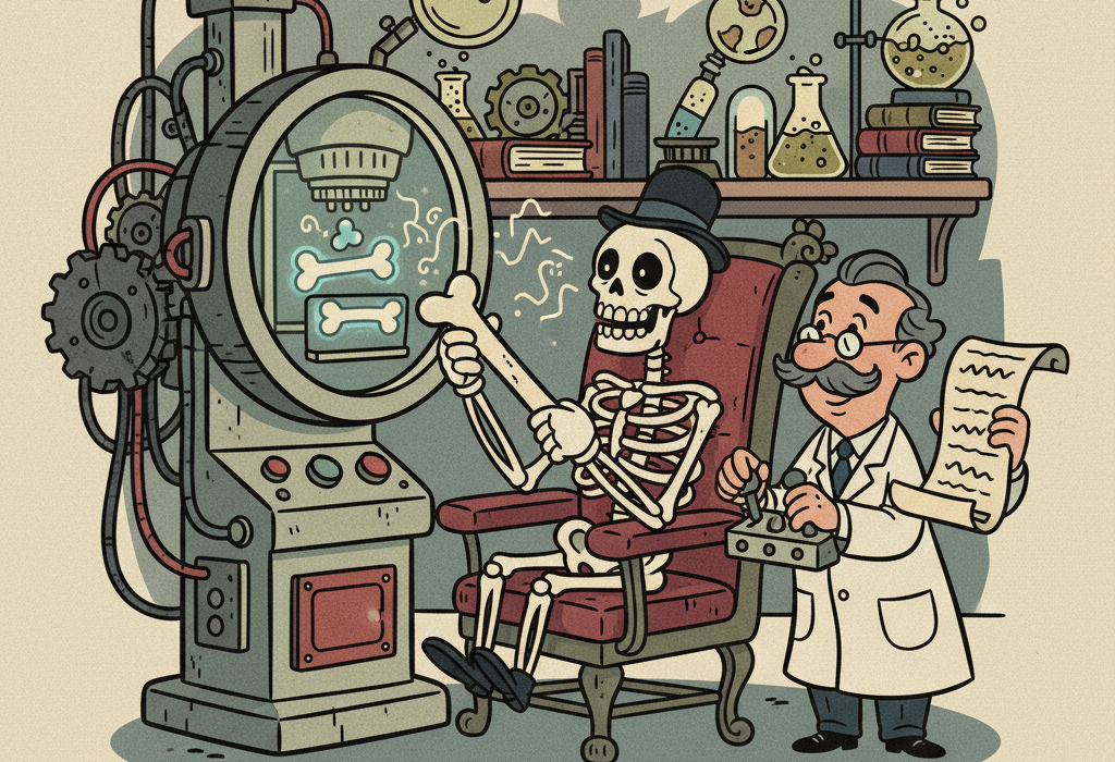
The gold standard for measuring bone health is a test called dual-energy X-ray absorptiometry (ab-sorp-shee-OM-eh-tree), commonly known as a DXA or DEXA scan.
A clinical review in Arquivos Brasileiros de Endocrinologia e Metabologia explains that a DXA scan uses two low-dose X-ray beams to measure the density of your bones, usually at the hip and lower spine. Bone absorbs more X-ray energy than soft tissue. By measuring how much energy passes through, the machine calculates your bone mineral density.
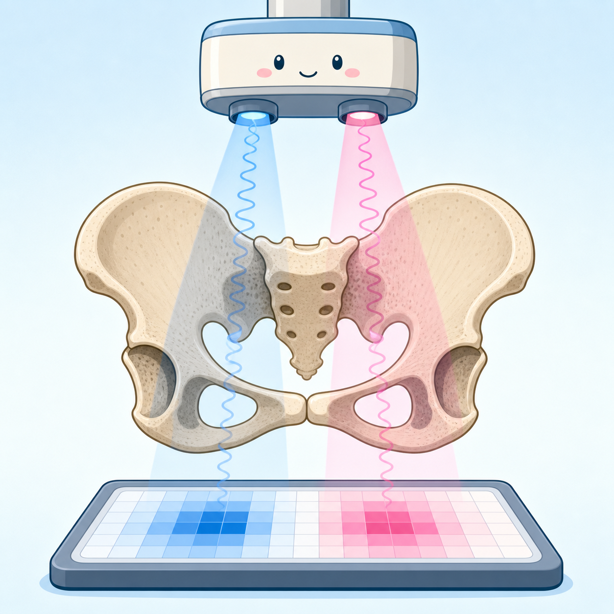
Science shows that bone is incredibly complex. A biomechanics study demonstrated that the hard outer shell of the bone (cortical bone) and the spongy inside (trabecular bone) actually behave as two completely different materials when put under stress. A DXA scan helps evaluate the overall structural integrity of these combined materials to predict how much force the bone can handle before breaking.
The Missing Diagnosis Problem
Despite clear guidelines, researchers have identified a widespread “care gap.” Millions of people who are at high risk for future fractures are not getting tested.
A large Medicare study in The Journal of Hand Surgery looked at older adults who broke a forearm or wrist. These types of breaks are major warning signs of weak bones. Yet, the researchers found that only 26% of these patients received a bone density test in the two years following their fracture.
Similar patterns exist worldwide. A study of patients in Japan found that less than 20% of people who suffered a hip fracture received a follow-up bone density test. Furthermore, a review in the Turkish Journal of Medical Sciences noted that up to two-thirds of spinal compression fractures go undiagnosed because patients assume the pain is just a normal part of aging.
Understanding Your T-Score
When you get a DXA scan, your results are usually given as a T-score.
A research update in Current Osteoporosis Reports explains that a T-score compares your bone density to that of a healthy young adult. Every one-point drop in your T-score roughly doubles your risk of a fracture.
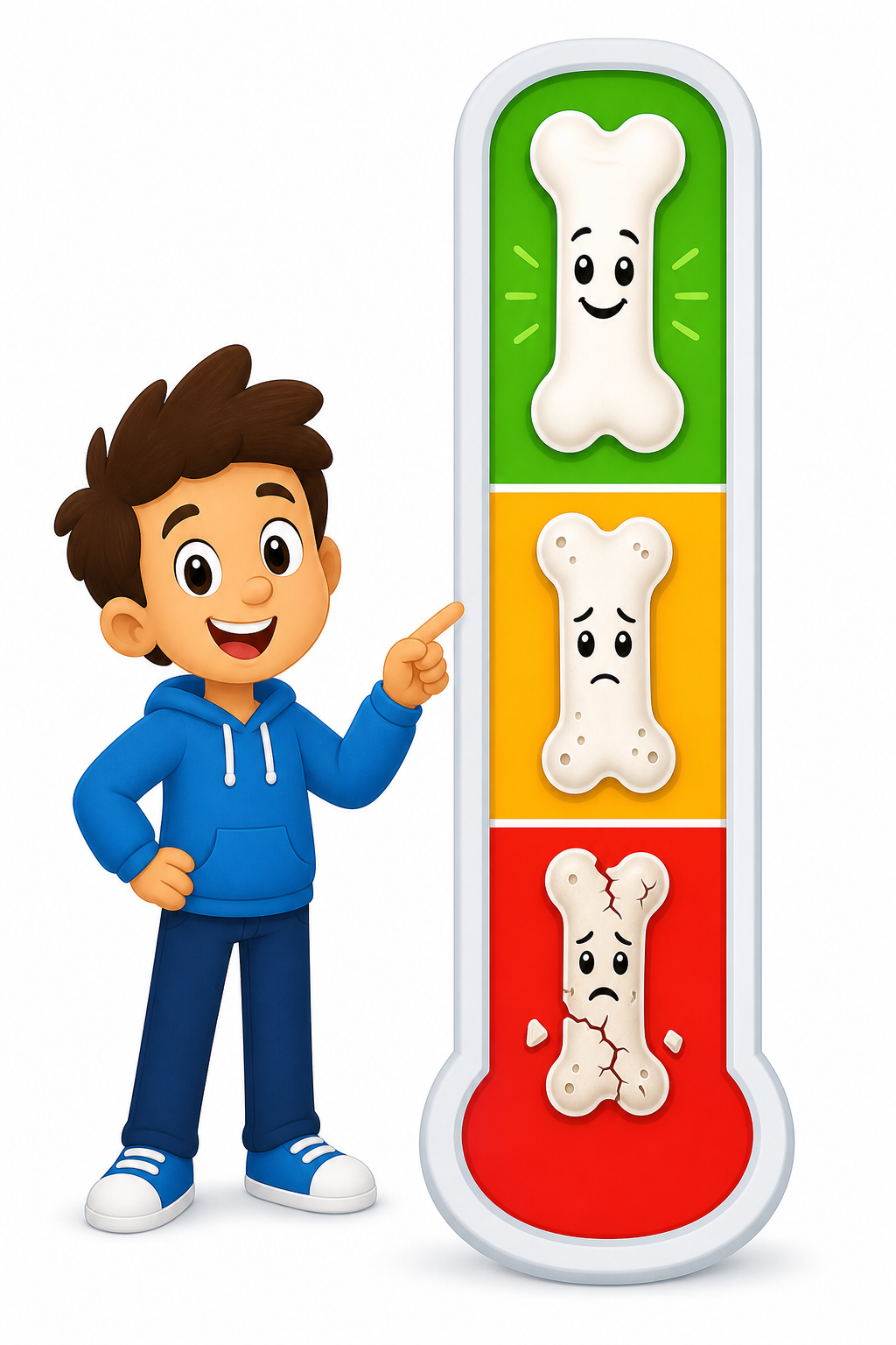
The World Health Organization classifies bone health into three main categories based on T-scores:
| Classification | T-Score Range | What It Means |
|---|---|---|
| Normal | -1.0 or higher | Bone density is within a healthy range. |
| Osteopenia | Between -1.0 and -2.5 | Bone density is low, but not yet in the danger zone. |
| Osteoporosis | -2.5 or lower | Bones are significantly weakened and at high risk of breaking. |
How Often Do You Actually Need a Scan?
A common question is whether you need a bone density test every year. For most people, the answer is no. Bone changes very slowly, typically losing only about 1% of density per year in older age.
Research shows that how long you should wait between scans depends heavily on your first T-score.
A study tracking rheumatoid arthritis patients and another study on thyroid cancer patients both found that the time it takes to develop osteoporosis varies greatly based on baseline bone health. Based on these findings, researchers suggest the following general intervals for repeat testing:
- Normal bone density: Wait 7 to 9 years.
- Mild osteopenia: Wait 5 to 7 years.
- Moderate osteopenia: Wait 3 to 4 years.
- Severe osteopenia: Wait 1 to 2 years.
Additionally, a study in the Journal of Bone and Mineral Research concluded that for women around age 65 with stable risk factors, waiting 5 to 6 years between scans is a safe and logical timeline.
Who Needs to Pay Extra Attention
While screening is generally recommended for all women over age 65 and men over age 70, certain groups need to be especially proactive about bone density testing.
People Who Have Broken a Bone: If you break a bone from a standing height (like tripping on a rug), this is called a fragility fracture. Research consistently shows that a prior fracture is one of the strongest predictors of a future fracture.
Men on Prostate Cancer Treatments: Some medical treatments actively drain bone density. A study in the journal Cancer looked at prostate cancer survivors receiving androgen deprivation therapy (ADT), which lowers testosterone and causes rapid bone loss. The researchers found that only 10% of these men received a bone density test, leaving a highly vulnerable group unprotected against fractures.
People Taking Steroids: Long-term use of glucocorticoids (like prednisone) can rapidly weaken bones, making early and frequent testing necessary.
Related: How to Prevent Age-Related Muscle Loss: What the Latest Science Says
Common Questions About Bone Density Testing
Does a bone density scan expose me to a lot of radiation?
No. A clinical review notes that the radiation exposure from a standard DXA scan is very low. It is roughly equivalent to the natural background radiation you experience during a regular day on Earth, or taking a cross-country airplane flight.
If my bones do not hurt, does that mean they are healthy?
Not necessarily. Osteoporosis is often called a “silent disease” because you cannot feel your bones getting weaker. The first symptom is usually a broken bone.
Are peripheral scans at the pharmacy just as good?
Devices that measure bone density at your heel, finger, or wrist are good for raising awareness, but they are not used to formally diagnose osteoporosis. If a peripheral scan shows low bone mass, doctors will typically order a central DXA scan of your hip and spine to confirm.
Related: The Science of Weight Training: How Resistance Exercise Changes Your Body
The Bottom Line
Bone density testing is a highly accurate, non-invasive way to measure the structural foundation of your body.
We know that DXA scans are the gold standard for predicting fracture risk. We also know that testing is vastly underutilized, especially for people who have already suffered a minor bone break or who take medications that weaken bones.
If your bones are currently healthy, you likely do not need a scan every year. But if you are over 65, have had a recent fracture, or have other risk factors, talking to your doctor about a baseline DXA scan could be the key to preventing a life-altering injury down the road.
Quick Reference: Key Studies
| Study Focus | Key Finding | Source |
|---|---|---|
| Medicare Patients & Fractures | Only 26% of older adults who broke a wrist/forearm received a follow-up bone density test. | PMID 33495040 |
| Testing Intervals | Patients with normal bone density can wait up to 7-9 years between scans, while those with severe osteopenia should test every 1-2 years. | PMID 27509834 |
| Prostate Cancer & Bone Loss | Only 10.2% of men on bone-depleting prostate cancer therapies received a recommended bone density test. | PMID 23065626 |
| Clinical Practice Updates | DXA scans remain the gold standard for diagnosis, and every 1-point drop in T-score roughly doubles fracture risk. | PMID 16303113 |
| Spinal Fractures | Up to two-thirds of spinal compression fractures go undiagnosed, highlighting the need for better screening. | PMID 32967415 |
Last updated: June 2026
This article synthesizes findings from peer-reviewed research. It is for educational purposes only and does not constitute medical advice. Consult a healthcare provider before starting any new regimen or if you have concerns about your bone health.

Leave a Reply