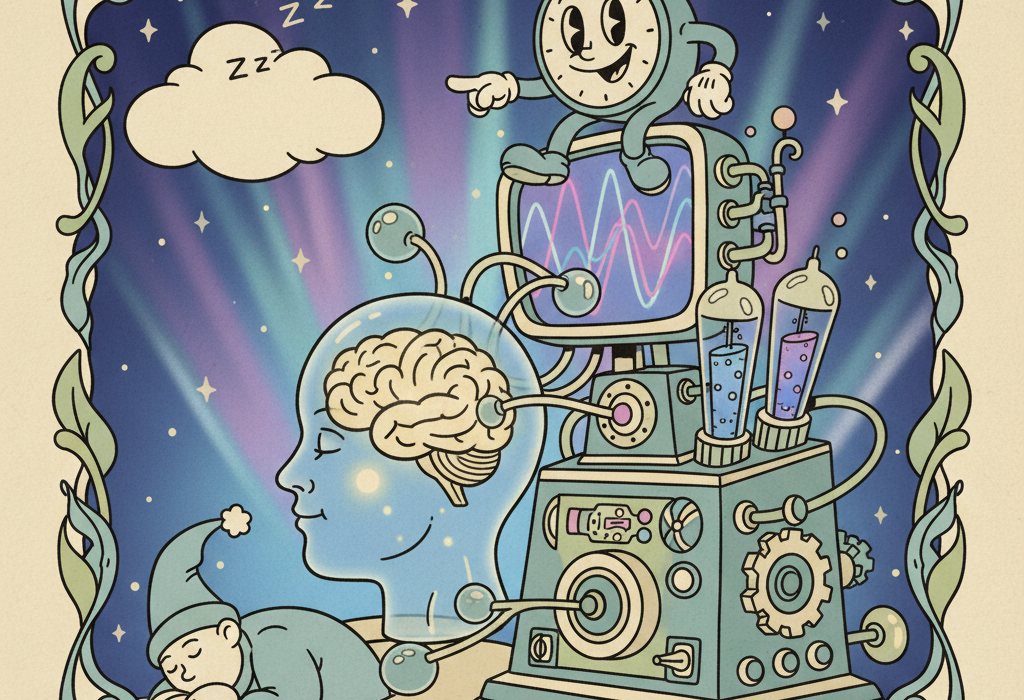
For decades, scientists could only study sleep by looking at brain waves on a screen. While this told us a lot about the different stages of sleep, it left a major question unanswered: what is actually happening to the physical structure and chemistry of the brain while we rest?
Today, advanced brain scans are giving us a literal look inside the sleeping brain. By combining traditional sleep tests with magnetic resonance imaging (MRI) and positron emission tomography (PET) scans, researchers can watch the brain clean itself, rewire its connections, and respond to sleep disorders in real time.
This article explores what the latest neuroimaging research actually shows about how sleep impacts brain aging, memory, and overall health.
How Science Looks Inside the Sleeping Brain
To understand the research, it helps to know how scientists gather this data. Sleep neuroimaging usually combines two methods:
- Polysomnography (pol-ee-som-NOG-rah-fee) – a comprehensive test that records brain waves, oxygen levels, heart rate, and breathing during sleep.
- Neuroimaging – techniques like MRI that show brain structure and blood flow, or PET scans that track how the brain uses energy and chemicals.
Running these tests together is notoriously difficult. As noted in a 2025 review in the Journal of Sleep Research, sleeping inside an MRI machine is loud, restrictive, and uncomfortable. Because of this, sleep imaging studies often have smaller sample sizes than other medical research. However, the data they do provide gives us a highly detailed picture of the brain’s nighttime activity.
What the Research Shows
Recent imaging studies have focused heavily on how the brain clears waste, how breathing disorders cause physical damage, and how poor sleep interacts with other health conditions.
The Brain’s Nightly Wash Cycle
One of the most important discoveries in modern sleep science is the glymphatic system (glim-FAT-ik) – the brain’s unique waste clearance pathway. During deep sleep, cerebrospinal fluid washes through the brain tissue, flushing out toxic proteins that build up during waking hours.
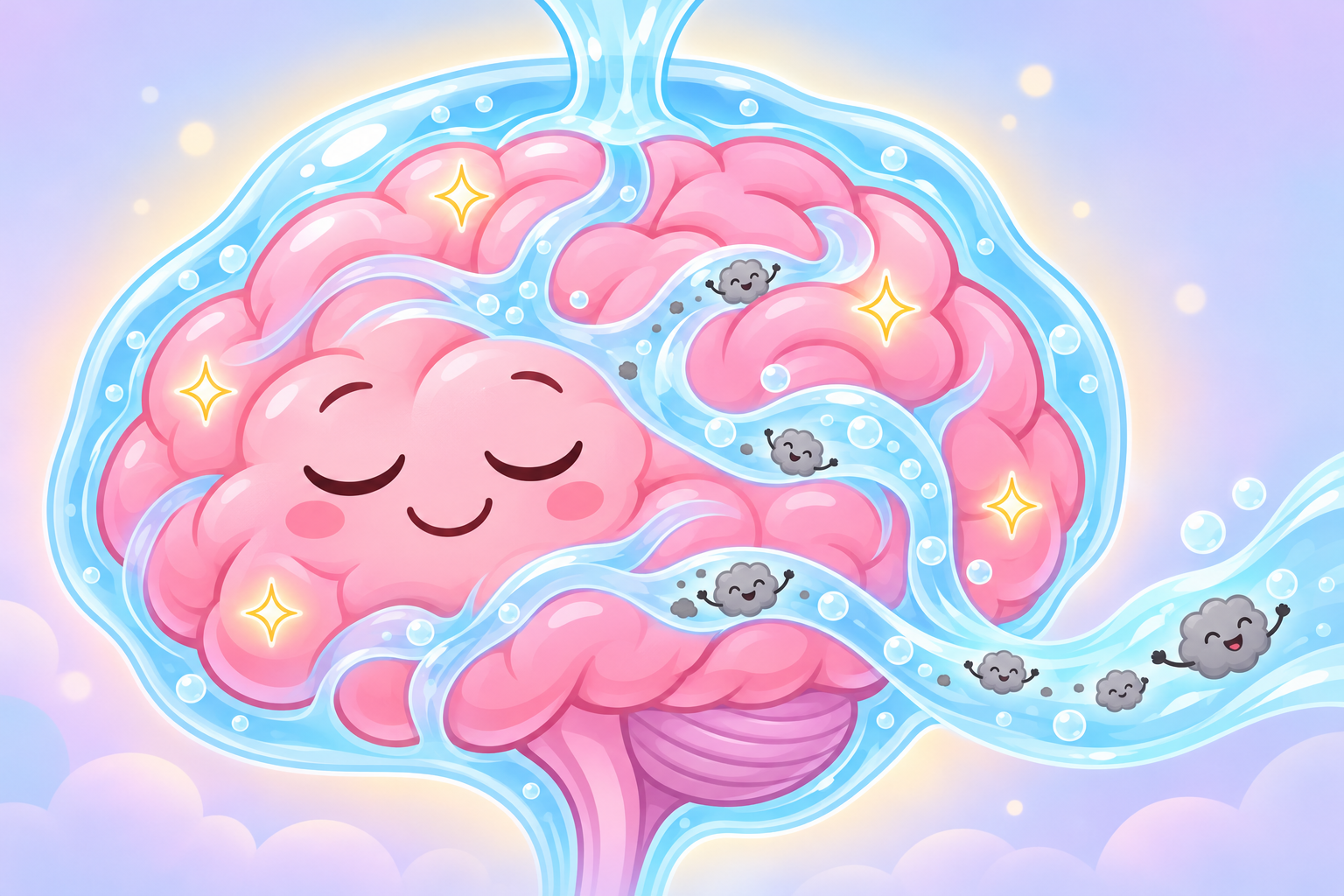
Scientists can now measure how well this system works using a specialized MRI technique called DTI-ALPS, which tracks the movement of water molecules in the brain.
A 2025 study in Molecular Psychiatry looked at 72 older adults and found that poor sleep quality directly correlated with a sluggish glymphatic system. The researchers discovered that when the brain’s washing mechanism slowed down, the structural connections between different brain regions weakened. This disruption was directly linked to memory decline in poor sleepers.
This cleaning system also appears to be an early warning sign for future neurological issues. A 2025 multi-center study in Neurology examined patients with isolated REM sleep behavior disorder (iRBD), a condition where people physically act out their dreams. The researchers found that these patients had significantly reduced glymphatic function compared to healthy adults. More importantly, those with the lowest fluid clearance were at a much higher risk of developing Parkinson’s disease years later.
The Toll of Sleep Apnea on Brain Structure
Obstructive sleep apnea (OSA) is a condition where the airway repeatedly collapses during sleep, causing temporary pauses in breathing. These pauses deprive the brain of oxygen and constantly pull the nervous system out of deep sleep.
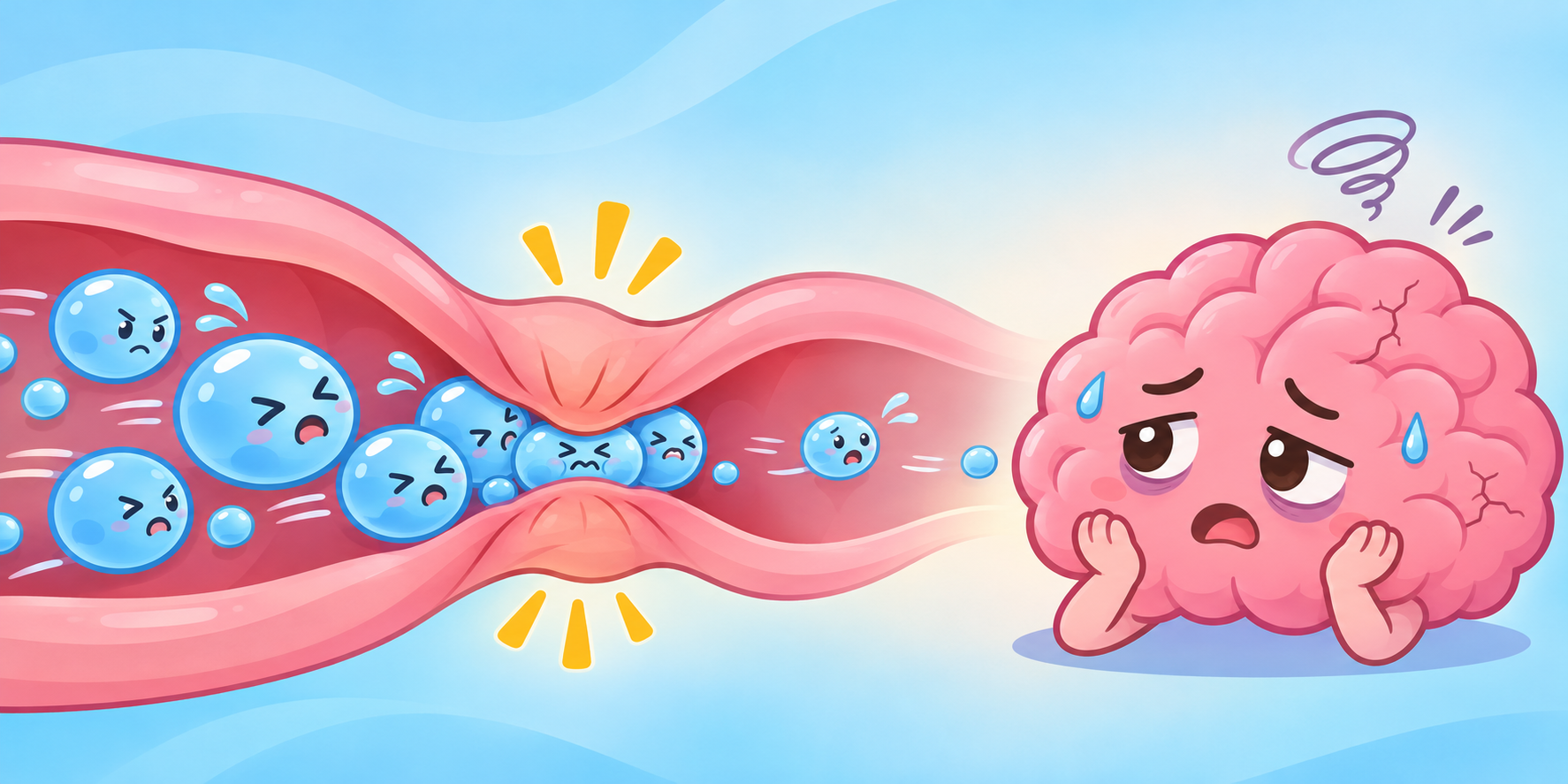
Brain scans show that this chronic lack of oxygen leaves a physical mark on the brain.
A 2021 study in Sleep analyzed MRI data from 690 adults. They found that the severity of sleep apnea was directly linked to “advanced brain aging.” The brains of people with untreated OSA looked structurally older than their actual age. The researchers noted that this aging process seemed to be driven by subclinical inflammation, evidenced by elevated white blood cell counts.
This damage is particularly concerning for those already at risk for cognitive decline. A 2026 study in the Journal of the Neurological Sciences looked at Alzheimer’s patients who also had sleep apnea. The researchers measured “hypoxic burden” (the total amount of oxygen deprivation during the night). They found that a higher hypoxic burden directly correlated with severe shrinkage in the left hippocampus and amygdala, which are the brain areas responsible for memory and emotional processing.
Interestingly, the brain’s response to sleep apnea is not always simple shrinkage. A 2025 study in BMC Medical Imaging found that people with severe OSA actually showed an increase in gray matter volume in certain areas. Researchers believe this is not a sign of health, but rather an adaptive, inflammatory swelling in response to chronic oxygen deprivation before permanent damage sets in.
Related: Understanding Brain Fog and Age-Related Cognitive Decline
The “Double Hit” of Short Sleep and High Blood Pressure
We know that high blood pressure damages delicate blood vessels, and we know that short sleep prevents the brain from repairing itself. When these two factors combine, the brain takes a “double hit.”
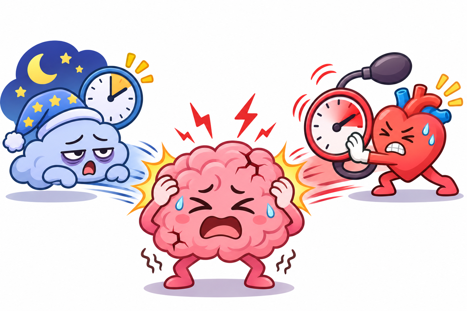
A 2024 study in the Journal of the American Heart Association examined 682 adults to see how blood pressure and sleep duration interact. They found that in people with normal blood pressure, sleeping less than six hours did not immediately show severe cognitive or structural brain damage.
However, in people with high blood pressure, short sleep was highly destructive. Hypertensive individuals who slept poorly showed worse executive function (decision-making and processing speed). Furthermore, their MRI scans revealed more “white matter hyperintensities” (small spots of vascular damage in the brain’s wiring) and increased shrinkage of subcortical gray matter.
This suggests that getting adequate sleep is especially critical for people managing cardiovascular conditions.
Related: How Sleep and Sleep Apnea Affect Your Heart Rate
REM Sleep, Mood, and Brain Networks
Rapid Eye Movement (REM) sleep is the stage where most of our vivid dreaming occurs. It is also deeply connected to our emotional health and mood regulation.
A 2025 study in Progress in Neuro-Psychopharmacology & Biological Psychiatry looked at 131 patients with major depressive disorder. Using functional MRI (fMRI), they found that patients with depression had less REM sleep and took longer to enter the REM stage. These sleep disruptions correlated with poor connectivity in the brain’s inferior parietal lobe, a region involved in emotional processing. When the patients’ depression symptoms improved over time, their brain connectivity also stabilized, highlighting the deep link between REM sleep and mood.
Brain scans also show how the brain tries to compensate when REM sleep goes wrong. In people who act out their dreams (iRBD), a 2019 PET imaging study in Sleep Medicine found unusually high levels of cholinergic activity in the brainstem. The researchers suggest that as the brain begins to experience early neurodegeneration, it temporarily pumps out more chemical signals to try and maintain normal sleep paralysis, before eventually failing as the disease progresses.
How This Might Work
The changes we see on brain scans generally come down to three biological mechanisms:
1. Oxygen Deprivation (Hypoxia): When breathing stops during sleep apnea, blood oxygen drops. Brain cells are highly sensitive to oxygen loss. Over time, this repeated stress causes cells to die, leading to the brain shrinkage (atrophy) seen on MRI scans.
2. Waste Buildup: The brain lacks a traditional lymphatic system. Instead, it relies on the physical shrinking of brain cells during deep sleep to allow fluid to wash away metabolic waste. If sleep is fragmented, this wash cycle is interrupted, leaving behind proteins that can damage neural networks.
3. Vascular Stress: Sleep is supposed to be a time when blood pressure dips, giving blood vessels a rest. Fragmented sleep keeps the nervous system on high alert, maintaining high blood pressure that slowly damages the tiny microvessels deep within the brain’s white matter.
Who Benefits Or Needs Caution
Based on the neuroimaging data, certain populations need to be particularly cautious about their sleep quality:
- People with hypertension: Because high blood pressure and short sleep compound each other to damage brain tissue, prioritizing sleep is vital for vascular health.
- People who snore heavily or gasp for air: These are hallmark signs of sleep apnea. The brain scans clearly show that untreated apnea accelerates brain aging and memory loss.
- Older adults experiencing memory issues: Ensuring proper deep sleep may help maximize the brain’s waste clearance system, potentially slowing cognitive decline.
Common Questions About Sleep and Brain Scans
Does sleep apnea permanently damage the brain?
Research shows that untreated sleep apnea leads to structural brain changes, including gray matter loss and increased signs of brain aging. However, other studies suggest that treating apnea with a CPAP machine can halt this progression and sometimes allow the brain to partially recover.
Why do scientists use both EEG and MRI for sleep studies?
An EEG (brain wave monitor) is excellent at telling scientists exactly what down-to-the-second stage of sleep a person is in. An MRI is excellent at showing exactly where blood is flowing and how brain structures look. Using them together allows researchers to see what physical parts of the brain are working during specific sleep stages.
Can a brain scan diagnose a sleep disorder?
Currently, no. Brain scans are used in research to understand how sleep affects the brain over time. Standard sleep disorders are diagnosed using polysomnography (overnight sleep studies) that measure breathing, heart rate, and brain waves.
The Bottom Line
Advanced neuroimaging has proven that sleep is not just a passive state of rest, but an active period of neurological maintenance.
We know with high confidence that poor sleep disrupts the brain’s waste clearance system and that oxygen deprivation from sleep apnea physically alters brain structure and accelerates brain aging. We also know that the combination of short sleep and high blood pressure is particularly damaging to the brain’s white matter.
While we still have much to learn about exactly how different brain networks communicate during REM sleep, the current science is clear: protecting your sleep quality is one of the most effective ways to protect your brain’s physical structure as you age.
Quick Reference: Key Studies
| Study Focus | Key Finding | Source |
|---|---|---|
| Glymphatic System & Memory | Poor sleep reduces brain waste clearance, directly correlating with memory decline in older adults. | PMID 39397082 |
| REM Sleep Behavior Disorder | Reduced brain waste clearance predicts future progression to Parkinson’s disease. | PMID 40956987 |
| Sleep Apnea & Brain Aging | Sleep apnea severity is linked to advanced structural brain aging and inflammation. | PMID 33017007 |
| Sleep Apnea & Alzheimer’s | Hypoxic burden from sleep apnea correlates with shrinkage in the hippocampus and amygdala. | PMID 42470846 |
| Short Sleep & Hypertension | The combination of high blood pressure and short sleep accelerates white matter damage and cognitive decline. | PMID 39450742 |
| Depression & REM Sleep | REM sleep disruptions in depression correlate with altered connectivity in emotional processing centers. | PMID 39326695 |
Last updated: November 2026
This article synthesizes findings from peer-reviewed research. It is for educational purposes only and does not constitute medical advice. Consult a healthcare provider before starting any new regimen.
Leave a Reply