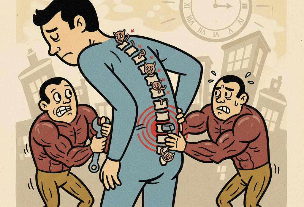
Anyone who has experienced a stiff, aching back knows how frustrating it can be to move through daily life. When your back hurts, your instinct is often to rest. However, researchers have discovered that prolonged back pain does more than just cause discomfort. It physically alters the muscles that support your spine.
For years, scientists and physical therapists have debated a core tension in spine health: does a bad back cause weak muscles, or do weak muscles cause a bad back?
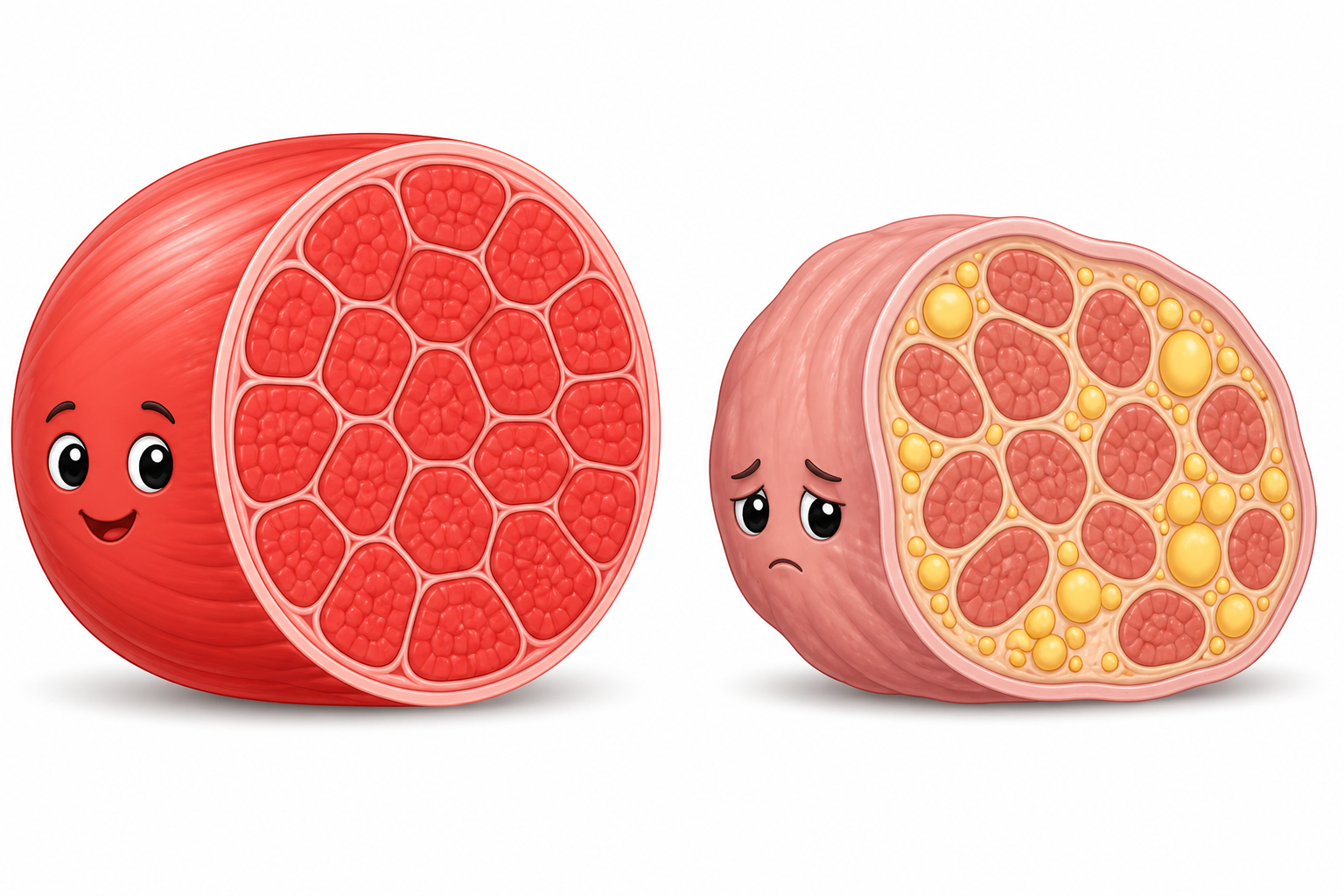
Modern imaging technology has finally provided some clear answers. Chronic low back pain is strongly linked to physical changes in the paraspinal muscles, specifically a decrease in muscle size and an increase in fat stored inside the muscle tissue.
This article breaks down what peer-reviewed research actually shows about how back pain changes your muscles, why it happens, and what the evidence says about reversing these changes.
Understanding Your Back Muscles
To understand the research, it helps to know the main players in your back. The paraspinal muscles (pair-uh-SPY-nul MUS-uhls) are the groups of muscles running down alongside your spine. They help you stand upright, bend, and twist.
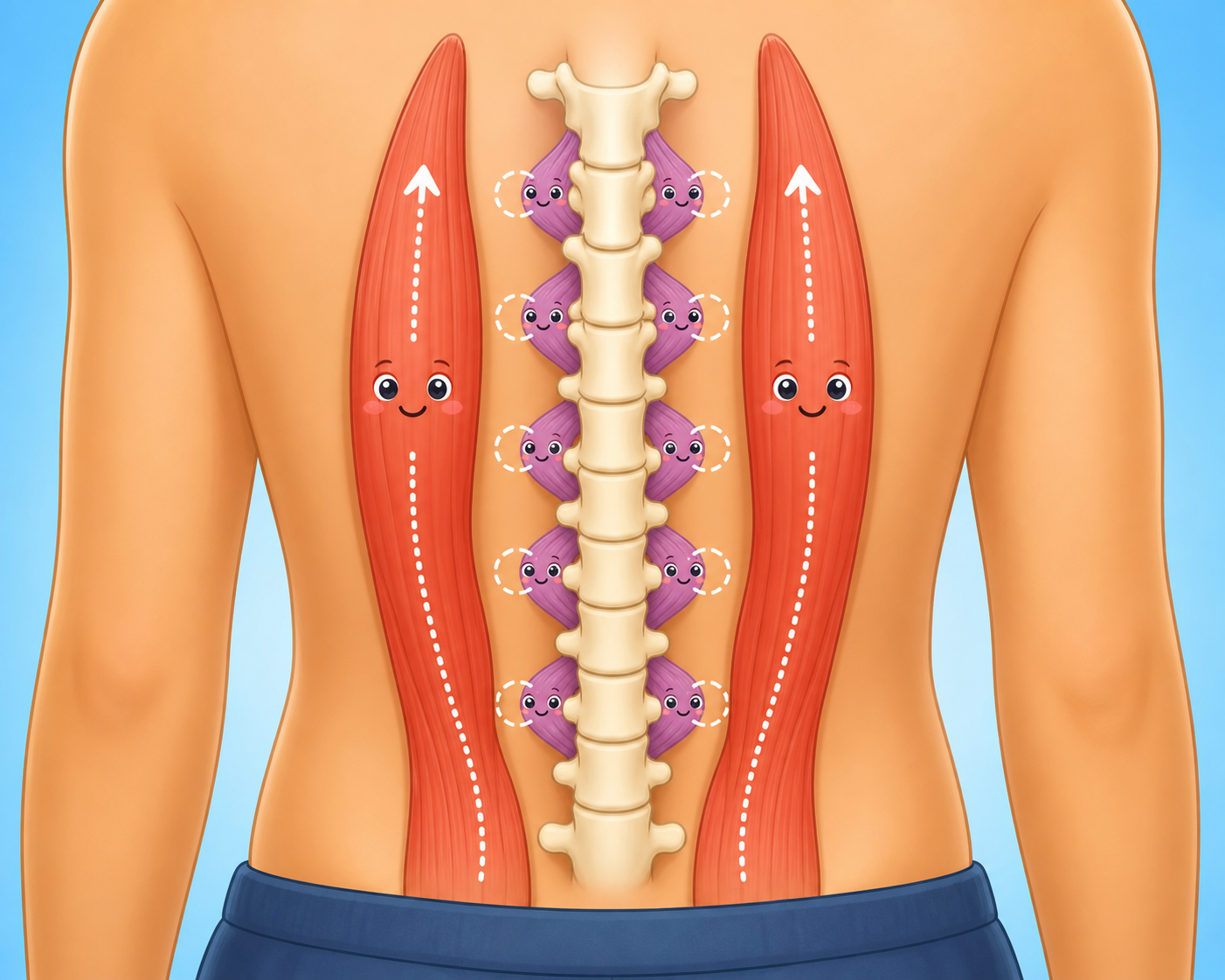
The two most studied paraspinal muscles in back pain research are:
- Multifidus (mul-TIF-ih-dus): A deep muscle that attaches directly to the spine. It acts like a series of small stabilizing cables, providing fine-tuned control over your spinal joints.
- Erector spinae (eh-REK-ter SPY-nee): A larger, more superficial group of muscles that primarily helps you straighten your back and lift heavy objects.
When scientists study these muscles using MRI or ultrasound, they look at two main measurements. The first is the cross-sectional area (KRAWS sek-shun-ul AIR-ee-uh), which is simply the overall size or thickness of the muscle. The second is fatty infiltration (FAT-ee in-fil-TRAY-shun), which occurs when healthy muscle fibers are replaced by fat cells.
What the Research Shows: The Anatomy of Back Pain
When you experience chronic low back pain, your body adapts. Unfortunately, these adaptations are not always helpful.
Muscle Shrinkage and Fat Accumulation
A massive 2024 study in The Spine Journal analyzed MRI data from 9,564 participants in the UK Biobank. The researchers found that people with chronic low back pain had significantly smaller paraspinal muscles and higher levels of fatty infiltration compared to people without pain. These changes were not just isolated to one small spot but were broadly distributed across the lumbar spine.
This finding is supported by a 2017 systematic review in The Spine Journal that looked at 25 different studies. The review found consistent evidence that a smaller cross-sectional area of the multifidus muscle is negatively associated with low back pain. In fact, a smaller multifidus muscle could predict future episodes of back pain up to 12 months in advance for men.
Interestingly, the evidence is less clear for the other back muscles. While the multifidus clearly shrinks in response to chronic pain, studies on the erector spinae and other surrounding muscles show mixed results. A 2016 systematic review in Pain Physician confirmed that muscle atrophy (shrinkage) in chronic low back pain is mostly localized to the multifidus muscle, while acute (short-term) back pain does not show these same structural changes.
Where the Fat Accumulates
Fatty infiltration does not happen evenly. A 2022 study in the European Spine Journal mapped exactly where fat accumulates in the muscles of people with chronic low back pain. The researchers discovered that fat tends to pool in the deepest regions of the multifidus muscle, right next to the center of the spinal discs.
This suggests a localized reaction. When a specific spinal disc is painful or degenerating, the deepest muscle fibers right next to that disc seem to shut down, eventually leading to fat replacing the inactive muscle tissue.
How This Might Work: The Biology of Muscle Changes
Why does a painful spine cause muscles to shrink and fill with fat? The answer involves a mix of biology, nervous system changes, and biomechanics.
Inflammation and Disc Degeneration
Spinal disc degeneration is a common cause of back pain. A 2022 study in Frontiers in Endocrinology used both human MRI data and a rat model to explore this connection. When researchers induced disc degeneration in the rats, they observed a direct increase in fatty infiltration in the nearby paraspinal muscles.
They also found elevated levels of a specific inflammatory marker called TNF-alpha in both the damaged discs and the surrounding muscles. This suggests that localized inflammation from a spinal injury might literally spill over into the muscle, triggering changes in how the muscle stores fat.
Brain and Nervous System Changes
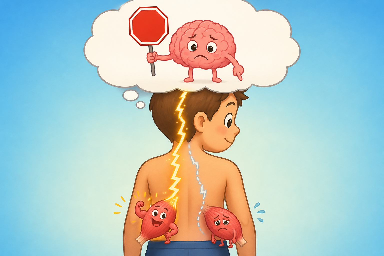
Pain also changes how your brain talks to your muscles. This is known as reflex inhibition. When a joint hurts, the nervous system reduces the electrical signals sent to the muscles around that joint to prevent further movement and potential damage.
A fascinating 2024 study in the Archives of Physical Medicine and Rehabilitation scanned the brains of patients with chronic low back pain while simultaneously measuring the electrical activity in their back muscles. The researchers found that abnormal spontaneous activity in the brain’s sensory and motor regions directly correlated with altered, inefficient activation of the lumbar paraspinal muscles.
Over time, this reduced communication means the muscle fibers are not being used. Just like a broken arm shrinking inside a cast, the unused back muscles begin to atrophy.
Reduced Oxygen and Early Fatigue
Because the muscles are smaller and less efficient, they have to work harder to do basic tasks. A 2024 study in Scientific Reports measured muscle oxygenation in people with and without chronic low back pain during a trunk extension exercise.
The researchers found that people with chronic pain had lower muscle oxygenation and reduced mechanical efficiency. Their muscles essentially ran out of gas faster. Another 2024 study in the Journal of Electromyography and Kinesiology confirmed this, showing that young adults with a history of back pain fatigued significantly faster during repeated endurance tests than those without a history of pain.
Can Exercise Reverse These Changes?
If pain causes muscles to shrink and fill with fat, can exercise reverse the process? The research says yes, though it requires targeted effort.
Core Stabilization vs. General Strengthening
Physical therapy is a frontline treatment for back pain, but researchers have debated which type of exercise is best.
Core stabilization exercises focus on retraining the brain to activate the deep multifidus and transversus abdominis muscles before moving the rest of the body. Strengthening exercises focus on building the larger, more superficial muscles like the erector spinae.
A 2021 randomized controlled trial in BMC Musculoskeletal Disorders compared these two approaches in patients with subacute low back pain. While both groups experienced pain relief, the core stabilization group saw significantly greater improvements in balance, proprioception (body awareness), and actual muscle thickness of the multifidus.
Similarly, a 2025 trial in BMC Musculoskeletal Disorders found that an 8-week core stabilization program yielded superior increases in multifidus cross-sectional area and more notable reductions in fat infiltration compared to conventional physiotherapy.
Related: The Science of Pilates: Benefits for Back Pain, Aging, and Beyond
The Overall Effect of Exercise Therapy
A comprehensive 2025 meta-analysis in the Annals of Physical and Rehabilitation Medicine looked at 34 different articles to determine the overall effect of exercise on muscle structure.
The researchers concluded that exercise therapy has a significant positive effect on increasing multifidus muscle thickness and cross-sectional area. The best results came from a combination approach: motor control training, stabilization, and lumbar strengthening combined.
However, it is important to note that a 2016 Cochrane Database Systematic Review found that while motor control exercise is very effective at reducing pain and disability compared to doing nothing, it is not necessarily superior to other forms of exercise or manual therapy in the long run. The best exercise is often the one a patient can perform consistently and safely.
Related: Strength Training for Back Pain: What the Latest Research Shows
Alternative and Complementary Therapies
While traditional land-based exercise is the most studied intervention, researchers have also looked at other ways to influence back muscle health.
- Aquatic Therapy: A 2025 study in Scientific Reports tested a 10-week aquatic exercise program against standard care. The water-based group saw significant increases in the volume of their multifidus and erector spinae muscles. Water therapy reduces spinal loading, making it a great option for people who find land-based exercise too painful.
- Dry Needling: A 2022 study in The Journal of Manual & Manipulative Therapy used ultrasound to measure muscle stiffness. They found that one session of dry needling reduced the resting stiffness of the erector spinae muscle after one week compared to a sham treatment.
- Restorative Neurostimulation: A 2025 review in Pain Practice highlighted restorative neurostimulation as a newer treatment for refractory cases. This involves an implanted device that stimulates the nerves controlling the multifidus muscle, essentially forcing it to contract and overriding the brain’s reflex inhibition.
Common Questions About Back Pain and Muscle Health
Does having a smaller back muscle mean I will definitely get back pain?
Not necessarily. While a smaller multifidus is a risk factor, it does not guarantee pain. A 2024 study on firefighters found no significant differences in back muscle strength or stiffness between those with and without a history of back pain, showing that highly active people can still experience pain without major structural deficits.
Can losing weight reduce the fat inside my back muscles?
While higher overall body mass index (BMI) is associated with more fat in the muscles, the fatty infiltration related to back pain is a localized response to spinal injury and muscle disuse. Losing weight is generally good for spine health, but specific targeted exercises are usually required to restore the deep stabilizing muscles.
How long does it take to rebuild the multifidus muscle?
Most clinical trials, such as those combining core stabilization and strengthening, show measurable improvements in muscle thickness and reductions in pain after 4 to 8 weeks of consistent exercise therapy.
The Bottom Line
Scientific research clearly shows that chronic low back pain is not just a sensation; it causes physical changes in your body.
- What we know: Chronic pain leads to the shrinking (atrophy) of the deep multifidus muscle and an increase in fat stored within the muscle tissue. This is likely caused by a combination of localized inflammation and the nervous system shutting down the muscle to protect the spine.
- What we know works: Targeted exercise therapy, specifically a combination of motor control, core stabilization, and strengthening, can successfully rebuild muscle thickness and improve function.
- What remains uncertain: While we know exercise helps, researchers are still debating whether specific core stabilization is vastly superior to general exercise in the long term, as consistency seems to be the most critical factor for pain relief.
If you are struggling with chronic low back pain, imaging might show that your muscles have weakened or accumulated fat. Fortunately, the evidence suggests that with the right rehabilitative approach, these muscles can be retrained and rebuilt.
Quick Reference: Key Studies
| Study Focus | Key Finding | Source |
|---|---|---|
| Muscle Changes in LBP | A study of 9,564 people found chronic LBP is associated with higher fat infiltration and lower muscle area across the lumbar spine. | PMID 38417587 |
| Core Stabilization | 8 weeks of core stabilization improved multifidus cross-sectional area and fat infiltration better than conventional physiotherapy. | PMID 41455992 |
| Exercise Meta-Analysis | A review of 34 studies found combining motor control, stabilization, and strengthening yields the best improvements in muscle structure. | PMID 40311262 |
| Brain and Muscle Link | Abnormal brain activity in chronic pain patients correlates directly with altered neuromuscular activation of the back muscles. | PMID 38969254 |
| Fat Distribution | Fat infiltration tends to pool in the deepest regions of the multifidus muscle, closest to degenerating spinal discs. | PMID 35776179 |
| Aquatic Exercise | 10 weeks of aquatic therapy significantly increased the volume of the multifidus and erector spinae muscles. | PMID 40328824 |
Last updated: November 2026
This article synthesizes findings from peer-reviewed research. It is for educational purposes only and does not constitute medical advice. Consult a healthcare provider before starting any new regimen.


Leave a Reply